


INSIGHTS TO PATHOPHYSIOLOGY AND RISK FACTORS OF CARDIAC ALLOGRAFT VASCULOPATHY: A PROSPECTIVE STUDY USING HIGHLY AUTOMATED CORONARY OPTICAL COHERENCE TOMOGRAPHY SEGMENTATION SOFTWARE IN 3D
Tématický okruh: Srdeční selhání, transplantace, oběhové podpory | |
| Typ: Ústní sdělení - lékařské , Číslo v programu: 211 | |
| Přihlášeno do: Soutěž mladých kardiologů | |
| Pazderník M.1, Bedáňová H.2, Kautzner J.1, Melenovský V.1, Karmazín V.1, Málek I.1, Tomášek A.2, Kovárník T.3, Chen Z.4, Šonka M.5 1 Klinika kardiologie, Institut klinické a experimentální medicíny- IKEM, Praha, 2 CKTCH, Brno, 3 II. interní klinika kardiologie a angiologie, VFN, Praha, 4 Institute for Biomedical Imaging, The University of Iowa, Iowa city, United States, 5 Institute for Biomedical Imaging, The University of Iowa, Iowa, United States | |
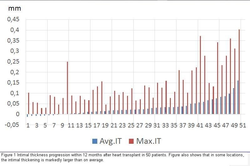
Introduction Current knowledge about cardiac allograft vasculopathy (CAV) is based mainly on data obtained from coronary angiography and intravascular ultrasound (IVUS) studies. Optical coherence tomography (OCT) is a new imaging modality that acquires images at a spatial resolution of 10-20 μm which is 10-fold better than that of IVUS. Methodology In a prospective, observational, multi-centre study consisting of 50 consecutive patients who underwent heart transplant (HTx) between 2014 and 2015 in IKEM Prague and CKTCH Brno. OCT imaging, presence of donor-specific-HLA antibodies (DSA), and 24-hour ECG Holter monitoring were analyzed in 1st and 12th months after HTx. Highly automated in-house developed segmentation software (IIBI, The University of Iowa) allowed quantitative analysis of intimal thickening over the entire 3D OCT pullback. Results 50 baseline (1 month post HTx) and follow-up (12 month) OCT pullback pairs from 50 HTx patients (407±69 overlapped image frames each, 20,341 frames total) were segmented and mutually registered. CAV manifested as a diasease with significant inter-individual progression variability. Average intimal thickness (IT) progression in the cohort ranged from -0.01 to +0.16 mm (Figure 1). Increased heart rate did not demonstrate as a risk factor for CAV development ( p= 0.65 at 1 month, p= 0.76 12 months after HTx). Analyzing presence of DSA did not show any association with average intimal thickness progression (p=0.47). Conclusion Using our automated OCT segmentation software, the reported work contributes unique data in heart transplantology: 1) CAV seems to be not only diffuse but also focal disease; 2) High proportion of patients develop severe progression of CAV already within 12 months after HTx; 3) Presence of DSA and increased heart rate are not associated with CAV progression. Supported by Grant 16-27465A | |
 |